 by
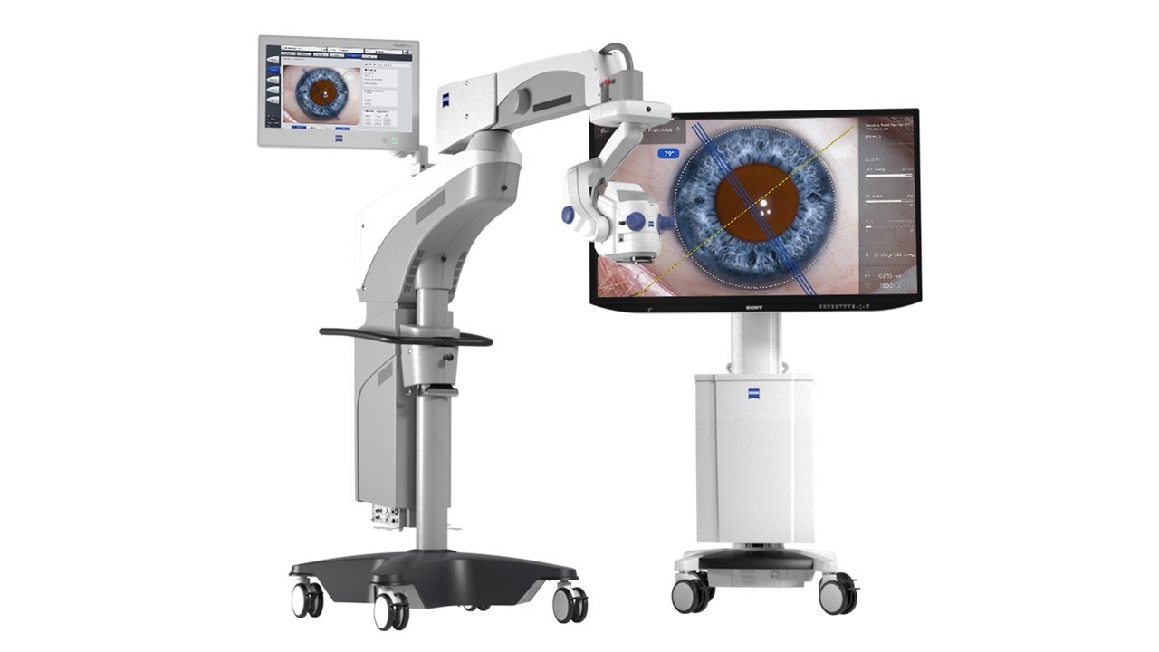
by Near infrared (NIR) medical imaging is an emerging non-invasive diagnostic technique that utilizes light in the near infrared range of the electromagnetic spectrum to visualize anatomical and physiological properties of tissues. By exploiting the ability of NIR light to penetrate deeper into biological tissues than visible light, NIR imaging has the potential to revolutionize areas like cancer detection, surgery guidance, and assessment of vascular health.
How Does NIR Imaging Work?
All objects above absolute zero emit electromagnetic radiation based on their temperature. Biological tissues are no exception and emit NIR light in the range of 700-900 nm. NIR imaging relies on this intrinsic tissue contrast to generate images. NIR cameras equipped with sensitive detectors capable of detecting this weak tissue-emitted light are used to acquire images. Some techniques may also employ NIR light sources to actively illuminate tissues and extract additional contrast. Unlike visible light, NIR light scatters and absorbs less in biological tissues, allowing it to penetrate deeper, up to a few centimeters below the skin surface. Specific tissues like hemoglobin, fat and water absorb NIR light differently based on their chemical composition, generating unique spectral absorption signatures that can be exploited for contrast. By analyzing these spectral absorption properties across the NIR range, rich physiological and molecular information about tissues can be derived.
Cancer Detection and Surgery Guidance
One of the most promising applications of Near Infrared Medical Imaging is for cancer detection and guidance during cancer surgeries. Many cancerous tissues have altered blood perfusion and angiogenesis compared to normal tissues, which results in unique absorption signatures in the NIR range. This property has enabled NIR imaging to detect cancers of the breast, colon, stomach and other organs with high sensitivity and specificity. During cancer surgeries, NIR cameras can help surgeons visualize the tumor margins in real-time and ensure complete removal of tumors along with the affected lymph nodes, thereby minimizing positive margin rates. Some studies have shown NIR imaging can reduce positive margins by over 50% compared to standard white light visualization alone for certain cancers. With its ability to assess cancer at the molecular level non-invasively, NIR imaging holds great potential to transform cancer diagnostics and improve outcomes.
Assessment of Vascular Health
Another important application of NIR imaging is for non-invasive assessment of vascular health. Hemoglobin is a strong absorber of NIR light and its oxygen saturation can be assessed using multispectral NIR images. This capability has enabled technologies like portable NIR finger clip devices to non-invasively monitor patients’ blood oxygen levels. NIR imaging techniques are also being developed to characterize microvasculature, blood perfusion and angiogenesis – all critical indicators of vascular health. Studies indicate NIR may help detect vascular diseases likePeripheral Arterial Disease at early stages by identifying alterations in blood perfusion patterns, years before symptoms appear. With an aging population and rising rates of vascular diseases worldwide, NIR imaging can play a big role in revolutionizing vascular health screening and management through its non-invasive, cost-effective nature.
Brain and Musculoskeletal Imaging
While most NIR imaging research has focused on oncology and vascular applications so far, its utility is not restricted to just soft tissues. Preliminary research indicates NIR may have applications in brain and musculoskeletal imaging as well. Due to its ability to penetrate the skull which blocks visible light, NIR imaging shows promise for non-invasive functional brain mapping and monitoring of patients with mild traumatic brain injuries. Studies have also demonstrated potential applications in cartilage imaging, with NIR contrast observed between healthy and degenerated cartilage tissues. Further research into these applications could uncover new diagnostic uses of NIR in neurology as well as orthopedics.
*Note:
1. Source: Coherent Market Insights, Public sources, Desk research
2. We have leveraged AI tools to mine information and compile it